Results for ""
Healthcare 4 Min Read Jan 11, 2024
Deep Learning model detects abnormal lung findings
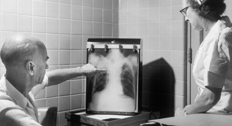
Artificial intelligence (AI)–assisted diagnosis imparts high accuracy to chest radiography (CXR) interpretation; however, its benefit for non-radiologist physicians in detecting lung lesions on CXR remains unclear.
Researchers allocated eligible patients who visited an outpatient clinic to the intervention (with AI-assisted interpretation) and control (without AI-assisted interpretation) groups. Lung lesions on CXR were recorded by seven non-radiologists with or without AI assistance. Three radiologists established the reference standard for lung lesions. The primary and secondary endpoints were the physicians’ diagnostic accuracy and clinical decisions.
An automated deep learning model accurately identified and predicted joint space narrowing and erosion based on hand radiographs of patients with rheumatoid arthritis, according to data presented at ACR Convergence 2023.
Between October 2020 and May 2021, 162 patients were assigned to the intervention and control groups. The area under the receiver operating characteristic curve was significantly more prominent in the intervention group than in the control group for the CXR and lung lesion levels. The intervention group had higher sensitivity regarding both CXR and lung lesion levels and a lower false referral rate for the lung lesion level. AI-assisted CXR interpretation did not affect the physicians’ clinical decisions.
The researchers found that AI-assisted CXR interpretation improves the diagnostic performance of non-radiologist physicians in detecting abnormal lung findings.
According to Carol Hitchon, MD, FRCPC, MSc, an associate professor at the University of Manitoba, said during a press conference, radiographic damage is one of the core outcome measures for rheumatoid arthritis, and as clinicians, we usually do serial plain X-rays so we can follow and detect joint damage in a patient over time, and that helps influence our management decisions.
One of the more commonly used metrics for evaluating joint damage, [the Sharp-Van der Heijde score], tells us how much collaborative space is narrowing and how many joint erosions there are — but it is not feasible in clinical practice.
She also stated that scoring these joints using these scores takes a lot of time. One needs a fair amount of expertise because there is a lot of intra- and inter-observer variability. Often, the expertise to do these scores and evaluate radiographs is unavailable in many centers.
Developing the system
To develop a deep learning system that would automatically detect joints and predict Sharp-Van der Heijde scores in patients with RA based on hand X-rays, Hitchon and colleagues first trained a convolutional neural network-based algorithm to see joints in 240 training and 89 test pediatric hand X-rays from the Radiologic Society of North America database.
Hitchon stated that they wanted to develop a model in which the computer can loot at and X-ray, and find a joint. The model will tell us what joint it is. It then detects if that joint is damaged and what the score for erosions and the score for joint space narrowing are.
Finding the results
Their results stated that the deep learning model could accurately identify the target joints, with a pediatric F1 score of 0.991 and adult data F1 score of 0.812. Predictions from the vision transformer model for erosion and joint space narrowing were highly accurate, with a root main squared error of 0.93 and 0.91, respectively.
Hitchon believes this model may have applications in larger studies such as randomized clinical trials or drug trials with large cohorts in which the outcome measure is joint damage.
Source: Healio Rheumatology